Glaucoma. Classification, treatment
GLAUCOMA - comes from the ancient Greek meaning
green, light blue.
Glaucoma is a serious disease that occurs throughout
world and usually affects people over 40 years of age, but
occasionally occurs at younger ages.
Glaucoma accounts for about 4% of all eye diseases. How
evidenced by the results of mass preventive
examinations, among the healthy population aged 40 years and
older, the disease occurs in 1-2% of cases.
In all countries of the world, glaucoma is one of the first
places as a cause of blindness.
The problem of successfully combating blindness from glaucoma is not
only the task of eye doctors, but also a general medical one
task. Therefore, doctors of all specialties should know
signs of this disease and how to treat glaucoma.
The duty of doctors of all specialties is to participate in
carrying out active preventive measures
facilitating early detection of the disease and treatment,
which prevents blindness from glaucoma.
Classification of glaucoma
primarysecondary
congenital
Primary glaucoma is one of the most common causes of irreversible blindness. In its development, there are 2 main pathophysiological mechanisms:
disruption of intraocular outflowfluid in the anterior part of the eye
apple;
optic atrophy
Direction of action of intraocular pressure
Open angle glaucoma (OAG)
The pathogenesis of open-angle glaucoma is associated with a violationfunctions of the drainage system of the eye, through which
drainage of fluid from the eye. On histological examination
eyes with OAG in the drainage zone of the limbus are always detected
dystrophic changes. In the initial stage of the disease
trabecular plates thicken and narrow
intratabecular clefts and especially the scleral sinus. IN
later the trabecula is completely degenerated, the gaps in it
disappear, the scleral sinus is overgrown. Last time
Evidence has accumulated that indicates an important role in
pathogenesis of OAG of the functional block of Schlemm's canal. All
these changes to a certain extent depend on the nervous,
endocrine and vascular disorders, therefore primary
Glaucoma is combined with diseases such as
atherosclerosis, hypertension, diabetes, lesion
subcutaneous region.
Both the anatomical features of the glaucomatous eye and
the nature and degree of dystrophic changes in the drainage
apparatus are determined by genetic factors, due to
than, primary open-angle glaucoma often carries
hereditary nature. Very often, OAG occurs and progresses unnoticed.
a patient who does not experience any unpleasant
sensations and consults a doctor when he notices significant
blurred vision. Usually normal due to some stagnation in
veins of the head due to the horizontal position of the body in
sleep time (in the morning) IOP is slightly increased, and by the end of the day it
decreases somewhat. The amplitude of P oscillations is not normally
exceeds 5 mm Hg. In glaucoma, these fluctuations
much bigger. The first and leading sign when
glaucoma is the presence of ophthalmotonus greater than 27 mm Hg.
Art., and fluctuations in the flow of the grids over 5 mm Hg. At
transition from the initial stage to the developed stage is marked by the second
a sign of glaucoma is a change in visual function,
consisting in a narrowing of the field of vision and a decrease in acuity
vision. Usually the narrowing of the visual field begins with the nasal
sides. The third cardinal sign of glaucoma is
expansion of the optic disc excision develops
in its later stages, due to expansion and protrusion
posteriorly, under the influence of increased IOP, the cribriform plate
and atrophy of nerve fibers and glial tissue. Fourth
sign - retinal edema, which is determined by
increasing the size of the blind spot.
Pseudoexfoliation glaucoma
Angle-closure glaucoma (ACG)
The main link in the pathogenesis of PACG is the blockadeangle of the anterior chamber with the root of the iris,
which arises as a result of functional
pupil block.
Functional or relative pupillary
block occurs in eyes with excessive anterior
location of the lens.
In such eyes, the iris is tightly adjacent to the front
surface of the lens, which impedes outflow
fluid from the posterior chamber to the anterior chamber. This
leads to increased pressure in the posterior chamber
eyes and protrusion of the iris anteriorly,
as a result, the angle of the anterior chamber
narrows, and under certain conditions the angle
closes. A specific role in the pathogenesis of PACG
belongs to genetic, nervous, endocrine
and vascular factors. Angle-closure glaucoma is common
(about 90% of all cases). This variety
glaucoma usually begins with acute or
subacute attack.
Diagnosis of PACG during acute and subacute
It’s not difficult to stage an attack. For early
PCG diagnostics use load
samples, of which the most effective and
safe dark and positional (face down).
The patient is placed in a dark room for 1 hour,
the sample is considered positive if
ophthalmotonus will not increase during this period
less than 5 mm Hg, and the position test
consists in placing the patient
on the couch face down also for 1 hour.
Increase in ophthalmotonus by 5 mmHg. Art. And
more indicates a predisposition to
blockade of the anterior chamber angle. Effect
dark test is associated with pupil dilation in
darkness, position test - with displacement
lens under the influence of gravity to the side
cornea.
Intraocular fluid flow in angle-closure glaucoma
Acute attack of glaucoma
Acute attack - occurs under the influence of various factors, withemotional stress, prolonged exposure to darkness,
medicinal dilation of the pupil or without any apparent reason.
The patient complains of pain in the eye and head, blurred vision,
the appearance of rainbow circles when looking at a light source. Painful
sensations are associated with compression of the nerve elements of the iris root
membrane and ciliary body.
With a severe attack, nausea and vomiting may occur.
The pain radiates to individual organs - the heart, the abdominal area, what
sometimes causes diagnostic errors. With objective
examination, stagnant injection of the vessels of the eye is striking
apple The cornea is swollen, like foggy glass, the anterior chamber
small (slit-like) pupil dilated.
Dilation of the pupil is associated with paresis of its sphincter caused by a sharp
increase in IOP. The iris is edematous, posterior synechiae are formed. Ocular fundus
visible in the fog:
The optic disc is swollen, with unclear contours. Often
you can see pulsation of the retinal artery and sometimes hemorrhages on the disc and
beside him.
During an acute attack, IOP rises to 70 mm Hg, outflow
fluid from the eye stops completely.
Gonioscopy reveals complete closure of the anterior chamber angle.
A subacute attack of glaucoma is characterized by the same basic
symptoms, but they are much less pronounced.
Accumulation of fluid behind the vitreous in malignant glaucoma - ciliary block
Differential diagnosis of an acute attack of glaucoma and acute iritis (iridocyclitis).
Acute attack of glaucoma Acute iritisaccompanied by complaints
(iridocyclitis). Rainbow
on rainbow circles at
there are no circles. Prevail
looking at the light.
pain in the eye. Eye
Prevail
falls ill suddenly.
radiating pain.
Prevails
Often preceded
pericorneal
prodromal seizures.
injection. Cornea
Congestive injection
transparent
vessels. Cornea
Sensitivity
diffusely cloudy.
the cornea is preserved.
Sensitivity
Anterior chamber depth
There is no cornea.
normal or
The anterior chamber is shallow.
uneven. Iris
The color of the iris is not changed,
hyperemic, altered
or changed
in color, the relief is smoothed.
insignificant. Pupil
The pupil is constricted. IOP is normal
wide, sharp IOP
or downgraded.
increased.
Classification of primary glaucoma
FormStage
State
IOP
Dynamics
visual
functions
Closed angle
Open angle
Mixed
Initial (I)
Developed (II)
Far
dropped by
(III)
Terminal (IV)
Normal
(A) T≤26mmHg
Moderately
increased
(B) T=26-32 mm
Stabilized
Unstabilized
Hg
High (C)
T≥33 mm Hg.
Acute attack of angle-closure glaucoma
Additional classification scheme for primary glaucoma
FormClosed angle
Variety
With pupillary block
Creeping
With a flat iris
With vitreocrystalline
block (malignant)
Main part location
outflow resistance
Pretrabecular
textile
Open Angle Simple
Pseudoexfoliative
Pigmented
Trabecular tissue
Intrascleral zone
(including collapse
Schlemm's canal)
Mixed
Combined
defeat
Clinical signs of the disease
In diagnosing the form of glaucoma, in addition to the clinical picture, gonioscopy is important - a method of examining the angle of the anterior chamber
For this you need to haveslit lamp and goniolens.
Three-mirror Goldmann lens. It is used for gonioscopy - examination of the angle of the anterior chamber.
Degree of opening of the anterior chamber angle (Grade 0 – angle closed, Grade 4 – angle open)
The degree of opening of the anterior chamber angle according to Shaffer
Gonioscopic picture in open-angle glaucoma
Gonioscopic picture of the anterior chamber angle in angle-closure glaucoma
Scheme of the gonioscopic picture in pigmentary glaucoma
Gonioscopic picture in pigmentary glaucoma
Anterior chamber angle in congenital glaucoma
Phacolytic glaucoma
I. Initial stage. At this stage of primaryno glaucoma, marginal excavation noted
ONH and pronounced changes in the visual field.
Dilatation may occur
physiological excavation of the optic disc, the appearance
small scotomas in the field of view (scotoma
Bjerrum - Fig A) and increase in size
blind spot.
II. Developed stage. For this stage
characterized by a persistent narrowing of the field of view by 100
from the nasal side and its concentric
narrowing There is a marginal excavation of the optic disc.
III. Far advanced stage. This stage
characterized by a persistent narrowing of the field
view from the nasal side or concentrically
up to 150 from the point of gaze fixation.
IV. The diagnosis of terminal glaucoma may
be installed in the complete absence
vision (Visus=0) or the presence of light perception with
incorrect projection of light (1/∞ l. incertae)
with at least partial transparency
avg.
Automatic perimetry
This is what glaucoma patients see
Advanced stage (II)Advanced stage (III) Optic nerve is normal
Glaucomatous
excavation Optic nerve is normal
Glaucomatous
excavation
Physiological excavation of the optic disc
Expansion of physiological excavation at the initial stage of glaucoma.
Marginal glaucomatous excavation in advanced stages of glaucoma
Tonometry is an objective method of measuring intraocular pressure
To assess intraocularpressure (IOP) is more often used 10g
Maklakov tonometer and accepted
the following gradations: A –
normal pressure (16 – 26 mm
Hg); B – moderately increased
(27-32 mmHg); C – high (33 and
above mmHg)
Congenital glaucoma
Congenitalglaucoma
Congenital iridocorneoendothelial syndrome
Classification of secondary glaucomas
1. Inflammatory andpost-inflammatory glaucoma:
a) caused by sclerites and
keratitis;
b) postveal;
c) with heterochromic uveopathy.
2. Phacogenic glaucoma:
a) phacotopic;
b) phacomorphic;
c) phacolytic.
3. Vascular glaucoma:
a) neovascular;
b) phlebohypertensive.
4. Dystrophic glaucoma:
a) with retinal detachment;
b) with iridocorneal
endothelial syndrome;
c) with primary systemic
amyloidosis;
d) hemolytic.
5. Traumatic glaucoma:
a) contusion;
b) wound;
c) burn;
d) radiation.
6. Postoperative glaucoma:
a) aphakic;
b) after keratoplasty;
c) after surgeries for detachment
retina.
7. Neoplastic glaucoma:
a) for intraocular tumors;
b) for orbital tumors and
endocrine exophthalmos.
Rubeosis of the iris. Secondary neovascular glaucoma
Phacolytic glaucoma
Conservative treatment
To reduce IOP, miotics are widely used, which are divided intocholinomimetic and antecholinesterase. Under the influence of miotics, iridescent
the membrane is pulled away from the angle of the anterior chamber, its rigidity increases and
bombing is decreasing. This mechanism is important in PACG. From
cholinomimetics use a solution of pilocarpine hydrochloride 1%, 2%, 4%,
carbocholine 0.75% – 3%. The disadvantage of cholinomimetics is their short duration
their action (4-6 hours).
The following anticholinesterose miotics are used in clinical practice:
actions: phosphakol 0.02%, armin 0.05%, 0.01%, fosarbine 0.01%, pibufine 0.025%,
tosmilen 0.1-1%.
All of these drugs are stronger than cholinomimetics. Their miotic action
lasts more than a day. Long-term use of anticholesterol miotics
action often causes the development of cataracts.
Sympathicotropic drugs. From this group of drugs for the treatment of glaucoma
Adrenaline, fethanol and euspiran are used. The mechanism of hypotensive action is associated
with improving the outflow of fluid from the eye and reducing the formation of watery
moisture. Adrenopilocarpine has a good effect. In recent years, widespread use
received β-blockers. These drugs lower IOP by suppressing the production of
aqueous moisture. Timolol 0.25% or 0.5% 1-2 times a day.
Carbonic anhydrase inhibitors - these drugs reduce the rate of formation
aqueous humor by 50%. The most widely used drug is acetazolamide.
called "diacarb". Prescribed orally 0.125-0.5 g 1-2 times a day (most often
O.25g is used 2-4 times).
Osmotic agents: urea 10% solution, mannitol IV 20% solution, ascorbate
sodium iv 20% solution, Glycerol orally.
TREATMENT OF ACUTE AND SUBACUTE ATTACK OF GLAUCOMA
Instillation of 1-2% is prescribed within 1 hourpilocarpine into the conjunctival sac every 15 minutes,
then every 1/2 hour, and then every hour (2-3 times).
After 6 hours, the frequency of instillation is reduced to 6 times per
day. Timolol is prescribed simultaneously with miotics
or clonidine (2-3 times a day), orally diacarb (0.5g,
then 0.25g 4 times a day), glycerin (1-2 times a day), i.m.
lasix, 2 hours hot foot baths. 2-3 leeches per
temple, after 3-4 hours IM chlorpromazine or lytic mixture
(aminazine, diphenhydramine, promedol). If in a day
the attack does not stop; iridectomy is indicated.
Intraocular fluid flow after iridectomy
Laser trabeculoplasty. Effective in the early stages of open-angle glaucoma. The procedure is painless and performed on an outpatient basis
conditions.Iridectomy is a fistulizing operation effective for closed-angle glaucoma.
Surgical iridectomyLaser iridectomy
Sclerectomy is a fistulizing operation in which an additional pathway for the outflow of intraocular fluid into the intrascleral veins is created.
Sclerectomy is a fistulizing operation in whichan additional pathway for the outflow of intraocular fluid is created
fluid into the intrascleral veins. Effective for many
types of glaucoma and has many modifications.
Slide 1
GRODNO STATE MEDICAL UNIVERSITY DEPARTMENT OF ENTRY, OPHTHALMOLOGY AND DENTISTRY LECTURE No. 5 Glaucoma. Clinic, diagnosis, treatment, prevention of glaucoma. Lecturer: Assoc. S. N. IlyinaSlide 2
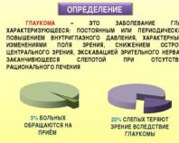
 GLAUCOMA IS AN EYE DISEASE CHARACTERIZED BY: CONSTANT OR PERIODIC INCREASE IN INTRAOCULAR PRESSURE, CHARACTERISTIC CHANGES IN THE VISUAL FIELD, REDUCED CENTRAL VISION ACUTUITY, EXCAVATION OF THE OPTIC NERVE AND ENDING BLINDNESS OH IN THE ABSENCE OF RATIONAL TREATMENT, 3% OF PATIENTS SEEK APPLICATIONS 20% OF BLIND PEOPLE LOSE VISION DUE TO GLAUCOMA
Slide 3

Slide 4
 I. COMPLAINTS FEELING OF PRESSURE IN THE EYES FEELING OF “TEARS”, FOREIGN BODY “BLACK FLOOSTERS” IN FRONT OF THE EYES PHOTOPSIES EARLIER PRESBYOPIA II. DETERMINATION OF IOP TONOOMETRY ACCORDING TO MAKLAKOV AND 24-hour tonometry ELASTOTONOMETRY PALPATORNO TONOGRAPHY III. STUDY OF THE ANGLE OF THE ANTERIOR CHAMBER GONIOSCOPY WURGAFT METHOD IV. STUDY OF PERIPHERAL VISION PERIMETRY CAMPIMETRICS V. BIOMICROSCOPY AND OPHTHALMOSCOPE
Slide 5

Slide 6

Slide 7

Slide 8
 I. PRIMARY 1. ACCORDING TO THE ANGLE OF THE ANTERIOR CHAMBER - OPEN-ANGLE - CLOSED-ANGLE - MIXED 2. BY STAGES OF DEVELOPMENT - INITIAL - DEVELOPED - ADVANCED - TERMINAL 3. ACCORDING TO IOP CONDITION - NORMAL (UP TO 27 mm) - MODERATE BUT INCREASED (28-32 mm) - HIGH (MORE 32 mm) IN THE DYNAMICS OF VISUAL FUNCTIONS - STABILIZED - UNSTABILIZED II. SECONDARY III. CONGENITAL IV. JUVENILE
Slide 9
 I. OPEN-ANGLE GLAUCOMA IS INDEPENDENT INTRAOCULAR PRESSURE IS INCREASED (NOT ALWAYS) PROGRESSIVE NARROWING OF THE VISUAL FIELD EXCAVATION OF THE OPTIC NERVE DISC OPEN ANGLE OF THE ANTERIOR CHAMBER REDUCED CENTRAL VISION II. CLOSED-ANGLE GLAUCOMA OCCUPIES IN PITCHY LAYS, THE EYE HURTS, REDUCED SENSITIVITY OF THE CORNIA, SWELLING OF THE CORNEA, CONSTANT INJECTION, SMALL ANTERIOR CHAMBER, ANTERIOR CHAMBER ANGLE IS CLOSED BY THE ROOT OF THE IRIS, COMPLAINTS ABOUT IRISIS GI
Slide 10
 I. OPEN-ANGLE GLAUCOMA - DRUG TREATMENT PILOCARPINE β-BLOCKERS (TIMOLOL, OPTIMOL, ARUTIMOL) TRAVATAN, XALATAN AZOPT, TRUSOPT ANTIOXIDANTS VASCODILATES TISSUE THERAPY - LASER TREATMENT - X SURGICAL TREATMENT II. CLOSED-ANGLE GLAUCOMA SURGICAL TREATMENT: SINUSTRABECULECTOMY
Slide 11
 I. COMPLAINTS RERADIATING HEADACHE REDUCED VISUAL ACUITY RAINBOW CIRCLES WHEN LOOKING AT A LIGHT SOURCE II. OBJECTIVE CONTAGENT INJECTION CORNEAL EDEMA SMALL ANTERIOR CHAMBER WIDE PUPILS IOP INCREASED TO 40-50 mm. rt. Art. III. TREATMENT: CONSERVATIVE UP TO 12-24 HOURS FROM THE MOMENT OF ATTACK. IF IOP DOES NOT DECREASE – IRIDECTOMY. TRABECULECTOMY IS PLANNED.
 Truth is an error that we temporarily believe to be correct. . .

 “Yellow-green water” (Russian)Grun. Star (German) Glaucoma (French, English) Jaskra (pol.) “Blakytna water” (Ukrainian)
 Glaukomas - the owl-eyed Heinrich Schliemann (Troy)
 HISTORY OF THE ISSUE Avicenna (980-1037), having begun to heal at the age of 18, described glaucoma a thousand years ago. . . at the turn of the 1st millennium In his “Canon of Medical Science,” republished in 1994, there is a description of an eye disease - “cold inflammation” associated with a violation of fluids in the body
 1000 years ago - Avicenna There is a type of inflammation of the eyes, which has periods and attacks, periods of change in matter and periods of time of its formation. The severity of pain during inflammation of the eye depends either on the burning juice, which corrodes the membranes, or on the abundance of juice, WHICH STRETCHES THEM. The matter that causes inflammation of the eyes comes either from the body in general, or from the head or from the vessels that bring bad matter to the eye, and sometimes the bad juices are in the eye itself.

 in the world: 7.8 million blind in both eyes with glaucoma. According to the International Society of Glaucomatology for 2008
 Incidence rates Statistical studies show that 1 in 200 over 40 years of age in the general population suffers from open-angle glaucoma. The overall prevalence of the population in this age group is 1.5%. The number of patients increases with age and reaches 12% in the group over 80 years of age.
 In the general population, primary OAG accounts for slightly less than 1%. Today in Russia there are more than 500 thousand patients with glaucoma, in the USA the number of patients with POAG is 2.47 million (out of a total population of 276.6 million people). According to the American Academy of Ophthalmologists (1996), 116 thousand Americans became blind as a result of glaucoma Incidence rates
 In the USA, 4% of the white population are blind in both eyes, 8% are black. Blind in one eye are 8% of the white population, 16% of the black population. The rate of blindness due to OAG in European countries averages 12% of all cases of blindness.

 PDA width options (Shaffer, Nesterov) 4 3 2 1 0 45 o 35 o 20 o 10 o
 Anatomical and physiological features of the drainage system of the eye The tissues of the drainage system are avascular Their metabolism is ensured by aqueous humor The trabecula contains an array of endothelial cells covering collagen fibers. In the cells, during metabolic processes, free radicals and lipid peroxidation products are formed that pass to the trabecula and damage it

 Etiological classification of glaucoma (D. Vaughan, T. Asbury, P. Riordan-Eva, 1999) A. Primary glaucoma 1. Vidkritokutova a. Primary glaucoma b. Glaucoma with normal (low) grip 2. Zakritokutova a. gostra b. podgostra v. chronic B. Congenital glaucoma 1. Primary congenital glaucoma 2. Congenital glaucoma is associated with another eye pathology 3. Congenital glaucoma is associated with an advanced congenital pathology C. Secondary glaucoma 1. Pigmented 2. Exfoliation syndrome 3. Phacogenic 4. Uveal 5 Iridocorneoendothelial syndrome 6. Traumatic 7. Postoperative 8. Neovascular 9. Advancing episcleral venous pressure 10. Steroid D. Absolute glaucoma Kintsevsky heritage of all species uncontrolled and glaucoma - important eye strain, blindness, often - pain
 Clinical classification developed by A.P. Nesterov and A.Ya. Bunin and adopted at the III All-Russian Congress of Ophthalmologists (1975). ACUTE ATTACK OF GLAUCOMA Form of glaucoma Stage IOP condition Dynamics of visual function Closed-angle Open-angle Initial I Developed II Unstabilized Stabilized. Normal (A) Moderately elevated (B) Far advanced III High (C) Mixed Terminal IV


 The hereditary form of congenital glaucoma is an autosomal recessive disease, which is based on underdevelopment of the drainage zone of the eye
 CAUSES OF CONGENITAL GLAUCOMA Unresolved embryonic tissue in the angle of the anterior chamber Anterior attachment of the iris root Underdevelopment of the trabecula Absence of Schlemow's canal Underdevelopment of intrascleral outflow tracts
 Clinical signs of congenital glaucoma Photophobia, blepharospasm, lacrimation Increase in the size of the cornea (from 9 to 22 mm) Edema, swelling, clouding of the cornea. Ruptures of Descemet's membrane Expansion of the limbus (from 1 mm to 3-4 mm) Staphylomas of the sclera Increase in the size of the eyeball (from 16 to 35 mm) Changes in the structures of the APC > IOP Glaucomatous excavation


 OBJECTIVE CHANGES IN OAG (in advanced and late stages of the disease) Emmysary symptom Cobra symptom Pathological pigmentation of the optic disc Dispersion of the iris pigment Atrophy of the iris stroma Leaching of the pigment border Presence of pseudoexfoliations Glaucomatous excavation of the optic disc Changes in the hydrodynamics of the eye Changes in the visual field
 Clinic of o/u glaucoma: Arises and progresses unnoticed Absence of pain and discomfort Complaints of periodic appearance of rainbow circles, blurred vision, sometimes headache in the superciliary area

 Risk factors for an acute attack of glaucoma Nervous tension Fatigue Staying in the dark Drug-induced mydriasis Prolonged head tilt Stress Taking large amounts of fluid
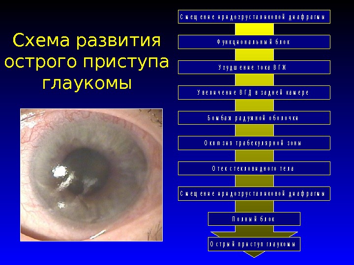 Scheme of the development of an acute attack of glaucoma. C h a r t T i t l e A c u c t u r e o f gla u co m a F u l l B l o c k S h i p t i v e i d i d o x p U s t a l d i a ph r a g m O f t e x t e x t o f t h e vitreous body o c l u s i o n a b e c u l a r y Z o n e B o m b a r i r iris I n c e l e r i n g I n g d R e a r c h a m m e r W e r d e r i n g i n g flow S h e n t i n g r i d o x r u s t i l a l d i a f r a g m
 links in the pathogenesis of glaucoma Specific atrophy of the optic nerve head with excavation. Hemodynamic disturbances in the optic nerve system OPTIC NEUROPATHY decreased perfusion Impaired autoregulation increased IOP apoptosis
 Control of IOP IOP - pressure of the contents of the eyeball on its walls Function of eye rigidity and its volume Depends on - production of intraocular fluid - outflow of intraocular fluid - volume of anatomical structures - resistance and blood supply of the vessels of the eye


 Volumetric model of normal and pathological differential light sensitivity as a function of localization in the visual field (Bebie. H., Fankhauser F., 1983) “Visual Island”


 Features of glaucomatous changes in the visual field Changes in the peripheral visual field in glaucoma. a - narrowing of the field of view on the nasal side, breakthrough of Bjerrum’s scotoma to the periphery; b - concentric narrowing; c - tube field of view; d - residual island of the visual field.
 In the first stage, a relative arcuate defect is detected. Isopter depression is often detected in the region from 5 to 25 degrees. from the fixation point, it is possible to identify a small scotoma in the same area. Five stages of development of glaucomatous visual field defects (Authorn, 1978) I
 In the second stage, the appearance of deep round-shaped defects, or scotomas, that do not merge with the blind spot is noted; they are often detected in the nasal region and an increase in the size of the blind spot. Five stages of development of glaucomatous visual field defects (Authorn, 1978) II
 In the third stage, an arcuate scotoma appears, often with a breakthrough to the periphery in the nasal region, which leads to the formation of a classic nasal step. Five stages of development of glaucomatous visual field defects (Authorn, 1978) III
 In the fourth stage, a widespread circular or semicircular scotoma may appear, leaving an island of vision in the center, as well as peripheral vision. Five stages of development of glaucomatous visual field defects (Authorn, 1978) I V
 In the fifth stage, the center of the optic hill practically collapses and only residual vision on the temporal side remains. Five stages of development of glaucomatous visual field defects (Authorn, 1978) V


 The number of capillaries in the layer of the cribriform plate is 3300. 1 bundle of axons is supplied with blood by 8 capillaries. There are 2500 capillaries in 1 mm of the optic nerve; 1 capillary supplies blood to 312 axons. Biological vulnerability of the optic nerve
 Biological vulnerability of the optic nerve absence of capillaries inside the axon bundles of the optic nerve insufficiency of blood supply to the axons decrease in the partial pressure of oxygen in post-capillary venules disproportion between the length of the axon (15 cm) and its thickness (15 mm)
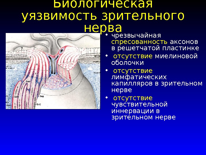 Biological vulnerability of the optic nerve extreme compression of axons in the cribriform plate absence of the myelin sheath absence of lymphatic capillaries in the optic nerve absence of sensory innervation in the optic nerve
 Anatomy of the blood supply to the optic nerve disc The main source is the posterior short ciliary arteries shown on the left side of the figure [according to Cioffi, van Buskrik, 1996].
 Quantitative criteria for the stage of glaucoma with a uniform expansion of the visible zone of axonal atrophy in all directions (type I excavation), if the E/D is:< 0, 4 — преглаукома; 0, 4 — 0, 5 — начальная глаукома; 0, 6 — 0, 7 — развитая глаукома; 0, 8 — 0, 9 — далеко зашедшая стадия. Достоверность показателей верифицируется меньшими размерами экскавации на парном глазу и наличием характерных для глаукомы нарушений зрения:

 Approximate forms for recording and sketching ophthalmoscopy data of the optic disc in the presence of signs of glaucoma are as follows. Example 1. Slight deflection of the temporal half of the optic disc of the left eye with an atrophy sector within one (inferotemporal) quadrant with moderate thinning of the disc with the presence of a beta zone in the disc atrophy sector with a width of about 0.1 (to the diameter of the disc). There are no hemorrhages.
 Approximate forms for recording and sketching ophthalmoscopy data of the optic disc in the presence of signs of glaucoma are as follows. Example 2. A barely noticeable total sagging of the optic disc of the right eye, emphasized by the course of the vessels, with a rounded zone of blanching in the center measuring E/D 0.7. In the 7 o’clock meridian there is a streak-like hemorrhage along the edge of the disc.
 Approximate forms for recording and sketching ophthalmoscopy data of the optic disc in the presence of signs of glaucoma are as follows. Example 3. The optic disc of the left eye in the entire temporal half with a transition below to part of the nasal half is grayish-white, excavated. The zones of sagging and blanching coincide. Over a long distance (in a sector of more than 6 hours), IU fishing is completely absent. The adjacent beta zone reaches a width of 0.3 (to the disc diameter).
 Approximate forms for recording and sketching ophthalmoscopy data of the optic disc in the presence of signs of glaucoma are as follows. Example 4. Total atrophic deep excavation of the optic disc of the right eye. Circular halo (Halo).

SSakh
Nuri Kumari
Chaudhary Suman
Lamichhane Sharmila
ml-402
GLAUCOMA
Glaucoma
Completed
Chaudhary Suman
Sah Nuri Kumari
Lamichhane Sharmila
Ml-402 DEFINITION
Glaucoma-
This
disease
eye,
characterized, characterized by: constant
or periodic increase in intraocular
pressure, characteristic changes in the visual field,
decrease
witticisms
central
vision,
excavation of the optic nerve and ending
blindness in the absence of rational treatment
:
3% OF PATIENTS
CONTACT
RECEPTION
20% OF BLIND PEOPLE LOSE
VISION RESULT
GLAUCOMA Character for them is
disturbances in the circulation of aqueous humor
(BB), leading to deterioration of its outflow from
eyes;
IOP is higher than tolerable for visual
nerve level;
ischemia and hypoxia of the ONH;
glaucomatous optic neuropathy;
degeneration (apoptosis) of ganglion cells
retina. Intraocular pressure
1.it ensures the maintenance of a spherical shape
eyeball and correct topographic
relationships between its internal structures,
2.facilitates metabolic processes in these structures
3.Influences circulation
blood in the intraocular vessels
*IOP level is relatively stable and varies
only in case of disturbances in the circulation of explosives. Circulation of aqueous humor
Moisture fills the posterior and anterior chambers of the eye and
flows mainly into the episcleral veins through the drainage
eye system located on the anterior wall of the angle
anterior chamber.
The EV first enters the posterior chamber of the eye and then through
the pupil passes into the anterior chamber, which serves as its
main reservoir.
With close contact of the iris with the lens, the transition
fluid from the posterior chamber to the anterior is difficult, which
leads to increased pressure in the posterior chamber Circulation of aqueous humor Anterior chamber angle (ACA) -
the narrowest part of the anterior chamber.
The anterior wall of the UPC is formed by a ring
Schwalbe, TA and scleral spur, posterior -
the root of the iris, the apex the base
ciliary crown Wide angle (40-45°) -
all structures of the Criminal Procedure Code (IV) are visible,
medium is not wide (25-35°) - determined
only part of the vertex of the corner (W),
narrow (15-20th) - ciliary body and glue
the ral spur is not visible (II),
visible slit (5-10°) - determined only
part TA (I),
closed - the structures of the Criminal Procedure Code are not
viewed (0).
Classification of the anterior chamber angle
eyes in width, a - wide; b -
medium width; c - narrow; G -
slit-like Drainage system
1. Trabecular apparatus – ring-shaped porous
crossbar between front and rear edges
internal scleral groove
2. Schlemm’s canal – scleral sinus located in
posterolateral part of the internal scleral groove
3. Outflow from the Schlemm canal through 20 -30 collector pipes
channels into the veins of the episclera CLASSIFICATION OF GLAUCOMA
1.
2.
3.
4.
I. Primary
According to the anterior chamber angle
Open angle
- Closed angle
Mixed
By stages of development
- Initial
- Developed
- Far gone
- Terminal
According to the IOP status
- NORMAL (UP TO 27 mm)
- MODERATELY INCREASED (28-32 mm)
- HIGH (OVER 32 mm)
According to the dynamics of visual functions
- Stabilized
- Unstabilized
II. Secondary
III. Congenital
IV. Juvenile closed angle glaucoma (ACG), with
in which the increase in IOP is caused by the milk block
UPC by intraocular structures
(iris, lens, glassy
body) or goniosynechia,
open angle glaucoma (OAG),
caused by damage to the drainage
eye systems,
mixed glaucoma, in which
Both mechanisms for increasing IOP are combined. Primary angle-closure glaucoma
Anatomical prerequisites –
1. small size of the eyeball;
large lens size;
anterior attachment of the iris to the CG
2. Age-related changes - flattening
cornea, iris atrophy in the root area. The pathogenesis of PACG is the closure of the UPC by the root of the iris.
The following mechanisms of such blockade are described.
As a result of the tight fit of the edge of the pupil to
lens, explosives accumulate in the posterior chamber of the eye, which
leads to protrusion of the iris root anteriorly and
blockade of the Criminal Procedure Code (Fig. 17.21).
The basal fold of the iris, formed when
dilation of the pupil, closes the filtration zone
narrow UPC in the absence of pupillary block.
Anterior displacement of the vitreous as a result of
accumulation of fluid in the posterior segment of the eye may
lead to the formation of vitreolenticular block.
In this case, the root of the iris is pressed against the lens
front wall of the UPC
As a result of the formation of adhesions (goniosynechia) and
root fusion
iris with the anterior wall of the UPC occurs
obliteration. Angle-closure glaucoma
1. It occurs in paroxysms, the eye hurts
2. Reduced sensitivity of the horn
shell
3.Swelling of the cornea
4.Congestive injection
5.Small anterior chamber
6.The angle of the anterior chamber is closed by the root
irises
7. Complaints about rainbow circles
8.High intraocular pressure (eye
dense as a stone)
9. Pupil dilation Acute attack of Glaucoma
A state of sudden and significant increase
intraocular pressure above 50 mmHg
Development mechanism
1.Impaired fluid circulation
2.Accumulation of excess fluid CLINIC
1. Blurred vision
2.Appearance of rainbows
circles around the source
Sveta
3. Sharp pain in the eye
4.iris bombing
5. Reduced severity
vision
6.Nausea
7.Vomiting
8.Dizziness Diagnosis of the disease
Superficial eye examination
redder than the eyes,
dilated oval pupil
no reaction to light)
1.
2. Upon palpation
increased tone
soreness An acute attack of glaucoma requires
emergency medical care
1. Miotics: pilocarpine 1-4% every 15 minutes for
1 hour, every hour during the day
2. ß – blockers 3 times a day
3. Diuretics:
Diacarb 0.25 – 4 times a day or IV
4.furosemide 40 mg
5. Distraction procedures:
leeches on the temple, mountains. Foot
baths
If the attack does not stop – after 12 – 24 hours
surgical treatment Open angle glaucoma
Anatomical prerequisites –
1.poor development of the scleral
spurs and ciliary muscles,
posterior attachment to sclera;
large lens size;
anterior attachment of the iris to
DH
2. Age-related changes in trabecular
apparatus, ciliary body, atrophy
irises
3. Genetic predisposition The pathogenesis of POAG includes three main
pathophysiological mechanism:
hydromechanical,
hemocirculatory and
metabolic.
The first one starts with
deterioration of the outflow of explosives from the eye and
increasing IOP.
Deterioration in outflow caused by
trabeculopathy - dystrophic
changes in TA. 2. An increase in IOP causes
decreased blood perfusion
pressure and intensity inside
ocular circulation, as well as
deformation of two mechanically weak
structures - the trabecular diaphragm in
drainage system of the eye and ethmoid
scleral plates.
3.Hemocirculatory disorders can be
divided into primary and secondary.
Primary disorders precede
increase in IOP, secondary ones arise in
as a result of the effect of increased IOP on
hemodynamics of the eye.
Clinic.
1.Complaints are absent or mild
2. Biomicroscopy: symptoms of “cobra” and “emissary”
3. Destruction of the pigment border
4.Pseudoexfoliation
5. Iris depigmentation
6. Intraocular pressure is increased (not always)
7. Progressive narrowing of the visual field
8. Excavation of the optic nerve head
9.Open angle of the anterior chamber
10Decreased central vision Primary open-angle glaucoma.
Gonioscopy.
1. Trabecular sclerosis
2. Pseudoexfoliation in the UPC
3. Deposition of pigment granules in
Code of Criminal Procedure DIAGNOSTICS
1.
2.
3.
4.
5.
1.
1.
2.
1.
2.
I. Complaints
Feeling of pressure in the eyes
Feeling of a “tear”, foreign body
“Black flies” before the eyes
Photopsias
Earlier presbyopia
II. Definition of IOP
Maklakov tonometry and daily tonometry
III. Study of the anterior chamber angle
Gonioscopy
Wurgaft method
3.Subjective assessment of the optic nerve disc condition
IV. Peripheral vision test
Perimetry
Campimetry
V. Biomicroscopy and ophthalmoscopy Increased IOP
1 Blockade of the RRU (corneal-iris
angle) root of the iris
2 Parietal blockade of foreign RRU
cloth
3Damage to the outflow tract
internal or external
walls of Schlemm's canal
4 Hypersecretion of EVs
IOP assessment in glaucoma
A - normal
20 – 26 mm Hg. Art.
B – moderately increased to
32 mmHg Art.
C – high 33 mm Hg. Art. And TONOMETRY ACCORDING TO MAKLAKOV Perimetry
NORM Early changes in visual field
Blind extension
spots
2.Scotoma in the area
Bjerrum (from 10 to
20°)
3. Seidel scotoma –
arched
scotoma in the area
Bjerrum
4. Rene's step
1.
Later changes
field of view
1.Ring-shaped
or double
arched
scotoma
2. Narrowing
nasal halves
3. Residual
central and
temporal
islets Late field changes
vision Excavation of the optic disc PROGRESSIVE LOSS OF VISUAL FIELDS IN GLAUCOMA.
Target -
1.reduce IOP by 30%
2.Influence microcirculation
in vessels
3. Improve nutrition of the MN and
retinas (neuroprotectors,
antioxidants) Drug treatment of glaucoma
1. Drugs affecting the outflow of intraocular fluid
2. Drugs that reduce production
VPG
3.Combined drugs The mechanism of their influence on IOP is related or
with improved outflow of explosives from the eye
1. Miotics
2.Adrenaline
3.latanoprost
decreased intraocular secretion
liquids
1.a2-adrenergic agonists,
2.p-adrenergic blockers,
3. carbonic anhydrase inhibitors. Cholinomimetics are used to reduce IOP
1. 1% Pilocarpine hydrochloride
2.pilocarpine hydrochloride
1% solution with methylcellulose
3. Carbocholine
4. Aceclin is used in the form of eye drops 3-6 times a day. Miotics cause contraction of the pupillary sphincter
and ciliary muscle,
promote dilation of blood vessels and
increasing their permeability.
Constricting the pupil and pulling the fold of the iris away from
UPC, miotics improve access of IVs to drainage
eye system
At the same time, due to the reduction of the ciliary
muscles stretch the trabecular diaphragm,
the blockade of Schlemm's canal decreases and
the outflow of explosives from the eye improves. Latanoprost (xalatan) - eye drops 0.005%
concentration - represents
synthetic analogue of prostaglandin F2o.
Latanoprost has a pronounced and
long-term hypotensive effect,
which is explained by the improvement of uveoscleral
outflow of B from the eye.
The drug is used 1 time per day. adrenergic stimulants in clinical practice
use
epinephrine dipivalate (dipivefrin) and
α2- adrenergic agonist (clonidine, clonidine). Carbonic anhydrase inhibitors
Acetazolamide
Dorzolamide hydrochloride Laser surgery for glaucoma. Laser
surgery is aimed primarily at
eliminating intraocular blocks along the way
movement of explosives from the posterior chamber of the eye into
episcleral veins. Laser iridetomy
consists in forming a small hole in
peripheral part of the iris.
The operation is indicated for functional or organic
pupil block.
It leads to equalization of pressure in the back and front
cameras of the eye and the opening of the Criminal Procedure Code. For preventive purposes
the operation is performed in all cases of angle-closure glaucoma
and with open-angle glaucoma with a narrow APC.
.Laser trabeculoplasty consists of
applying a series of cauterizations to the internal
surface of the trabecular diaphragm, in
as a result, its permeability to
BB and
the risk of blockade of Schlemm's canal is reduced.
The indication for surgery is POAG, not
compensable with
medicines Microsurgery of glaucoma.
Micro surgical operations are performed using
operating microscope and special microinstruments
cops
There is a wide variety of operational
interventions that can be divided into 4 main
groups..
Iridectomy
Fistulizing operations
trabeculectomy
Cyclodestructive operations Operations that improve the circulation of explosives inside
eyes, - iridectomy
(elimination of pupillary block) and
iridocycloretraction (expansion of the UPC).
The indication for performing these operations is
primary or secondary angle-closure glaucoma.
Fistulizing operations allow
create a new pathway for the outflow of IVs from the anterior chamber into
subconjunctival space, where fluid comes from
the bone is absorbed into the surrounding vessels.
(trabeculectomy) Non-Penetrating Filtration Operations (NPFO) are based on
on subscleral excision of the outer wall of the scleral
sinus (sinusotomy) in combination with trabecus stretching
polar wall using micro cauterization
According to one of the modifications of the operation (non-perforating
deep sclerectomy) deep limbo plate
scleral tissue is excised not only above Schlemm's
canal, but also anterior to Descemet’s membrane.
The effectiveness of NFO increases with the use of anti
metabolites during or after surgery. Decrease
severity of the hypotensive effect of NFO in
postoperative period serves as an indication for performing
laser perforation of the trabecular diaphragm in the area
operations Cyclodestructive operations are based on
damage and subsequent atrophy of part of the processes
ciliary muscle, which leads to
reduction in explosive production From modifications of this
neck operations are most widespread
cyclocryodestruction.
During the operation, several
cryoapplications on the sclera in the area of location
ciliary crown.
With sufficient intensity and for a long time
the strength of cryotherapy can be achieved significantly
reduction of IOP.
The length of the impact zone should not exceed
180-200° to avoid hypotension and atrophy transscleral diodelaser
cyclocoagulation,
characterized by greater safety and high
efficiency.
Cyclodestructive operations are indicated for far
advanced glaucoma, as an additional
intervention in case of unsuccessful outcome or incomplete
the effect of previously performed fistula-isolating
operations and for terminal glaucoma with painful syn
drome.
GBOU RM SPO (SSUZ)Saransk Medical College
Methodological development
for an open theoretical lesson on the topic:
« Violations of the hydrodynamics of the eye. Etiology, clinical manifestations, treatment. Emergency care for an acute attack of glaucoma"
Item - Eye diseases
Speciality - General Medicine
Qualification - paramedic
The level of education - increased
Completed by Rev. Shamshetdinova G.Kh.
Saransk 2012
Explanatory note
This lesson is devoted to the most pressing topic of our time, glaucoma. PAccording to literature data (including WHO), the number of glaucoma patients in the world reaches 100 million people. Glaucoma ranks first among the causes of visual disability.
The methodological development is intended for the training of paramedical workers in the specialty “General Medicine”, qualification “Paramedic” (advanced level of education) in the subject “Eye Diseases”.
This lesson is theoretical and is conducted in accordance with the lecture plan. To facilitate the perception of lecture material in class, multimedia illustrations are used.
During the lesson, in order to better perceive the material, fragments of techniques are used:critical thinking technology (compiling clusters and syncwines), which allowsorganizedtprocesstraining, aimed at the active activity of understanding, applying, analyzing, summarizing or evaluating information obtained or created through observation, experience, reflection, reasoning or communication as a guide to action or the formation of beliefs; case method allowingconsolidate the knowledge acquired in previous classes,develop skills in the practical use of conceptual schemes and familiarize students with schemes for analyzing practical situations, as well as develop skills in group problem analysis and decision-making (as part of training procedures);health saving technologies Anddevelopmental learning technologies .
To control and consolidate the acquired knowledge, situational tasks are carried out at the end of the lesson. Great importance is attached to the independent work of students; it was proposed to prepare short messages, reflecting their opinion on the topic under consideration.
Tasks:
1. To study the anatomical and physiological features and functions of the visual analyzer, their changes in glaucoma.
2.Study the research methods used in diagnosing glaucoma.
3.Study the clinical manifestations of glaucoma.
4. To study emergency care for acute and subacute attacks of glaucoma and the tactics of managing a patient with glaucoma.
5. Study the rules of dispensary observation of glaucoma patients.
Purpose of the lesson:
Educational - study the concept of etiopathogenesis, clinical manifestations, principles of treatment of glaucoma.
Developmental - promote the development of clinical thinking, memory, attention; instill the ability to work with educational and technical literature.
Educating - to promote the development of hard work, accuracy, a sense of responsibility and honesty, to influence the aesthetic views of students, to contribute to the formation of professional qualities in the work of a paramedic.
Interdisciplinary connections:
anatomy;
physiology;
therapy;
obstetrics;
pediatrics;
psychology.
Lesson timing:
1. Organizational moment: checking students’ attendance, checking readiness for the lesson, announcing the topic of the lesson, tasks and goals of the lesson, introductory word from the teacher - motivation for the lesson - 5 minutes.
2. Presentation of new material - 50 min.
2.1 Presentation of the material - 25 min.
2.2 Physical education minute - 3-5 min.
2.3 Presentation of the material - 20 min.
3. Student messages - 15 min.
4. Reinforcing the material (compiling clusters, syncwines, situational tasks) - 15 min.
5. Summing up - 5 min.________________________________
Total: 90 min.
Progress of the lesson.
1 Introductory word (5 minutes).
This lesson is devoted to a pressing topic of our time, namely the disease that ranks first among the causes of blindness and visual disability, glaucoma.Between 1% and 2% of the population over 40 years of age is affected by this disease.According to WHO, there are more than 100 million patients with glaucoma in the world, and their number is steadily increasing every year. According to the forecast, by 2030 this number could double.
Even at the level of modern knowledge, it remains unclear what triggers the occurrence of glaucoma. Many diseases inherent in old age contribute to its development. These include diseases of the cardiovascular system (atherosclerosis, hypertension), the endocrine system (diabetes mellitus, thyroid diseases), and the nervous system.
Hereditary factors play a huge role in the occurrence of glaucoma. It is 35%. Blood relatives of patients with glaucoma constitute the main risk group and should undergo an annual preventive eye examination.
2 Presentation of new material (50 minutes):
Glaucoma - a large group of eye diseases, each of which is characterized by a constant or periodic increase in intraocular pressure caused by impaired outflow of aqueous humor from the eye, resulting in the development of a special form of optic nerve atrophy with excavation in the disc area. Characterized by a triad of signs:
1 increase in intraocular pressure.
2 progressive glaucomatous optic atrophy.
3 progressive decrease in central and peripheral vision.
Etiological factors the occurrence of glaucoma: heredity, metabolic disorders, hemodynamic and endocrine disorders, local degenerative changes.
Classification:
By origin: primary, secondary and combined with developmental defects of the eye and other structures.
By patient age: congenital, infantile, juvenile and adult glaucoma.
According to the mechanism of increase in intraocular pressure: open-angle, closed-angle, dysgenesis of the anterior chamber angle, pretrabecular block and peripheral block.
By IOP level: hypertensive and normotensive.
According to the degree of damage to the optic nerve head: initial, developed, advanced and terminal.
Downstream: stable and unstable.
Pathogenesis associated with impaired outflow of aqueous humor - with the development of trabeculopathy and functional canalicular block, and is common to all forms of primary open-angle glaucoma. Violation of the hydrodynamics of the eye leads to an increase in IOP above the normal level and the development of optic disc atrophy of the glaucomatous type.
Congenital glaucoma.
The cause of congenital glaucoma is unresolved embryonic mesodermal tissue, anterior attachment of the iris, dysgenesis of the aqueous humor, leading to difficulty in the outflow of aqueous humor, and then to an increase in IOP.
Clinic. Congenital glaucoma is characterized by an inconspicuous course. You can suspect glaucoma based on mild photophobia, lacrimation, restless behavior, and the child’s sleep. The cornea, anterior chamber of the eye and pupil increase in size. The cornea may look like fogged glass or white porcelain. This appearance is most often caused by edema or dystrophy of the cornea.
Edema can be distinguished from dystrophy when one of the hypertonic solutions (glucose, urea, glycerin and even saline solution) is instilled onto the cornea: the edema disappears or decreases after instillation. But not only the size of the cornea can be increased to about 11 mm (and not 9 mm), the depth of the anterior chamber is 3-4 mm (and not 2 mm), the pupil width is 3-4 mm (and not 2 mm), but also the entire eyeball , and this increase reflects the stage of the glaucomatous process.
Enlargement of the entire eye is the so-called buphthalmos (bulls eye) or hydrophthalmos (dropsy of the eye). High ophthalmotonus is also evidenced by the presence of congestive eye injection with characteristic symptoms of “emissary”, “cobra’s head”, and processed “body of the medusa”. The magnitude of the increase in intraocular pressure is determined by palpation as T+2 and even higher (T+3).
If the cornea is transparent, then with ophthalmoscopy one can detect in the area of the optic nerve disc (papilla) a shift of the vascular bundle to the nasal side and pathological atrophic excavation of the disc itself.
All of the listed glaucomatous changes in various structures of the eye cannot but affect visual functions: they are reduced according to the stage of the process and the severity of symptoms.
Treatment: Surgical - goniotomy operation. The purpose of goniotomy is to destroy the mesodermal tissue in the angle of the anterior chamber and open the outflow of moisture from the anterior chamber through Schlemm's canal; or goniotonia with goniopuncture - + creation of additional outflow of intraocular fluid under the conjunctiva
If help is not provided, optic nerve atrophy will result; by one year the eye will go blind.
Open angle glaucoma accounts for more than 90% of all cases of this disease. With this form of glaucoma, the iridocorneal angle is open, which is why it gets its name. For reasons not yet fully understood, the outflow of intraocular fluid is disrupted. This leads to a buildup and a gradual but constant increase in pressure, which can ultimately destroy the optic nerve and cause vision loss.
In most casesopen angle glaucoma arises and progresses unnoticed by the patient, who does not experience any discomfort and consults a doctor already at a late stage of the disease, when he notices worseningvisual acuity. Complaints about the appearancerainbow circlesaround light sources, periodicblurred vision noted only by 15-20% of patients. These are the symptoms that appear whenand may be accompanied by pain in the eyebrow area and head.
Open angle glaucoma As a rule, it affects both eyes, in most cases occurring asymmetrically.
The leading symptom of the disease is increasedintraocular pressure (IOP) . Intraocular pressure at open angle glaucoma increases slowly and gradually as outflow resistance increasesintraocular fluid (IOH) . In the initial period it is unstable, then it becomes persistent.
The most important diagnostic signopen angle glaucoma is a changefield of view. First of all, these defects are determined in the central sections and are manifested by the expansion of the boundaries of the blind spot, the appearance of arched prolapses. These disorders are detected in the early stagesglaucoma, with special studiesfields of view. As a rule, patients themselves do not notice these changes in everyday life.
With further developmentglaucomatous process are revealed peripheral visual field defects . Narrowing field of viewoccurs predominantly on the nasal side, then the narrowing of the field of vision concentrically covers the peripheral parts until it is completely lost. Dark adaptation worsens. These symptoms appear against the background of a persistent increaseintraocular pressure (IOP) . A fall visual acuityspeaks of a severe, advanced stage of the disease, accompanied by almost completeoptic nerve atrophy .
Angle-closure glaucoma - a rarer form of glaucoma. Women get sick more often than men. Predisposing factors for the development of this formglaucoma are:
anatomical predisposition;
functional closure factorsanterior chamber angle ;
age-related changes in the eye.
Anatomical features of the structureeyeball , predisposing to the developmentangle-closure glaucoma serve small eye size, shallow anterior chamber, largelens , narrowanterior chamber angle , farsightedness . Functional factors include increased productionintraocular fluid (IOH) , increased blood supplyintraocular vessels , pupil dilation .
With this form of glaucoma, the pressure in the eye rises quickly. Anything that causes the pupil to dilate, such as dim light, certain medications, and even dilating eye drops given before an eye exam, can cause the iris to block the flow of fluid in the eye. When this form of the disease occurs, the eyeball quickly hardens and the sudden pressure causes pain and blurred vision.
Flowangle-closure glaucoma in most patients it is characterized by periodic, initially short-term, and then increasingly long periods of increaseintraocular pressure (IOP) . In the initial stage, this is due to mechanical closure of the zonetrabeculae iris root , which is due to the anatomical predispositions of the eye. Whereinoutflow of intraocular fluid (IOH) decreases. When fully closedanterior chamber angle a condition calledacute attack of angle-closure glaucoma . In the intervals between attacks, the angle opens.
During such attacks, adhesions gradually form between the iris and the wall of the anterior chamber angle, the disease gradually becomes chronic with a constant increase inintraocular pressure (IOP) .
Duringangle-closure glaucoma The following phases can be distinguished:
preglaucoma;
acute attack of glaucoma;
chronic course of glaucoma.
Preglaucoma occurs in individuals who do not have clinical manifestations of the disease, but upon examinationanterior chamber angle It turns out that it is either narrow or closed. Betweenpreglaucoma Andacute attack of glaucoma possible transient symptoms of visual discomfort, the appearance of rainbow circles when looking at a light source, short-termvision loss . Most often, these phenomena occur during prolonged exposure to darkness or emotional excitement (these conditions contribute to pupil dilation, which completely or partially reducesoutflow of intraocular fluid ) and usually disappear on their own, without causing much concern in patients.
Acute attack of glaucoma occurs under the influence of provoking factors, such as nervous tension, fatigue, prolonged exposure to darkness, drug-induced pupil dilation, prolonged work in a position with the head tilted, and intake of large amounts of liquid. Sometimes an attack appears for no apparent reason. The patient complains abouteye pain and in the head, especially in the back of the head,nausea, often vomiting, general weakness, blurred vision , appearance rainbow circles when looking at a light source. An acute attack of glaucoma is often mistaken for a migraine, hypertensive crisis, or poisoning, which leads to serious consequences, since such a patient must be helped in the first hours of the disease.
Painful sensations are caused by compression of nerve elements inroot of the iris Andciliary body . Visual discomfort is associated with corneal edema.
During a visual examination without special instruments, one can only notice a sharp dilation of the vessels on the anterior surface of the eyeball, the eye becomes “red”, somewhat with a bluish tint (congestive vascular injection). The cornea becomes cloudy due to the development of edema. Noteworthy is the dilated pupil that does not respond to light. At the height of the attack, it can sharply decreasevisual acuity . Intraocular pressure can increase to 60-80 mmHg. Art.,drainage of fluid from the eye stops almost completely. The eye feels dense to the touch, like stone.
If, within the next few hours after the onset of an attack, the pressure is not reduced using medications or surgery,eye faces irrevocablevision loss !!! Acute attack of glaucoma is an emergency and requires emergency medical attention!!!
Over time, the disease becomes chronic. This typeglaucoma occurs with a progressive riseintraocular pressure (IOP) , subacute attacks and increasing blockadeanterior chamber angle . These processes naturally end with the developmentglaucomatous optic atrophy , loss of visual function .
Urgent Care:
1 instillation of pilocarpine 1% for the first 2 hours every 15 minutes, then 2 hours. every 30 minutes, during track. 2 hours 1 time per hour. Then 3-6 times per day depending on the degree of IOP reduction.
2 0.5% timolol twice a day.
3 orally or parenterally osmotic diuretics, IM or IV furosemide 20-40 mg.
4 orally acetazolamide 0.25-0.5 g 2-3 times a day.
5 dorzolamide 2% 3 times a day.
6 IM lytic mixture: 1-2 ml 2.5% aminosine + 1 ml 2% diphenhydramine + 1 ml 2% promedol. After administration of the mixture, bed rest for 3-4 hours prevents orthostatic collapse.
7 to relieve an attack and prevent repeated attacks - laser iridectomy in both eyes.
8 if the attack could not be stopped within 12-24 hours, surgical treatment is indicated.
Secondary glaucoma is a complication of inflammatory diseases or injuries of the eye, as a result of which there is an outflow of fluid through the drainage system.
Stages of glaucoma:
Initial: the boundaries of the visual field are normal, but there are changes in the paracentral parts of the visual field (individual scotomas in zone 5-200 , Bjerum's arcuate scotoma, widening of the blind spot). M.b. initial signs of excavation of the optic nerve head.
Developed: pronounced changes in the visual field in the paracentral region combined with a narrowing of its peripheral boundaries by more than 100 in the upper or lower nasal segment. The excavation of the optic disc approaches the marginal one.
Far advanced: the border of the visual field is concentrically narrowed and in one or more segments is less than 150 from the point of fixation, there is a marginal excavation of the optic nerve head.
Terminal: complete loss of vision or preservation of light perception with incorrect light projection. The field of view cannot be examined.
IOP level: a-within normal limits (18-24 mm Hg); c-moderately elevated (25-32 mm Hg); c-high blood pressure (33 mm Hg).
Treatment:
1 first choice drugs: 1% pilocarpine, 0.25% and 0.5% timolol, 0.005% latanoprost. If the drug is ineffective, it is replaced with another drug of first choice or a combination of first and second choice (0.25% and 0.5% betaxolol, 1-2% proxodolol, 2% dorzolamide)
2 enzyme antioxidants - superoxide dismutase-Erisod.
3 1% emoxipine subconjunctival and parabulbar 10 injections. The effect of emoxipine is enhanced when combined with tocopherol. Aevit 1 capsule 2-3 times a day with vitamins gr. IN.
4 antispasmodics - orally xanthinol nicotinate 150 mg 3 times a day after meals for 2 months. or IM 15% 2 ml once a day for 10 days. Theophylline 250 mg 3 times a day for 2 weeks; Cavinton 5 mg 3 times a day for 1 month; pentoxifylline after meals 400 mg 3 times a day.
5 angioprotectors - dicinone 0.25 g 3 times a day for 2-3 months; prodectin0.25g 3-4r/d2-4wk
6 nootropic drugs - nootropil 30-160 mg/kg/day for 6-8 weeks. Picamilon 10 mg 3 times a day; antihypoxants (cytochrome C).
7 vitamin therapy.
8 piracetam - 30-160 mg/kg/day 6-8 weeks.
3.Student messages
Students give a presentation on the topic: “Dispensary observation of a glaucoma patient”- 15 minutes.
4. Reinforcing the material (compiling clusters and syncwines, situational tasks) - 15 min.
How to create a cluster
1. Write the keyword or sentence in the middle of a large piece of paper or on the board.
2. Write down words or sentences that come to mind about the topic.
3. As you come up with ideas, you write them down.
4. Write down as many ideas as come to your mind.
How to make a syncwine
Sinkwine is a “poem” consisting of five lines. In syncwine, a person expresses his attitude to the problem.
The order of writing a syncwine:
The first line is one keyword that defines the content of the syncwine.
The second line contains two adjectives that characterize this concept.
The third line is three verbs showing the action of the concept.
The fourth line is a short sentence in which the author expresses his attitude.
The fifth line is one word, usually a noun, through which a person expresses his feelings and associations associated with this concept.
Make a cluster for the word:
Glaucoma Congenital glaucoma
Disease
Decreased visual acuity
Limitation of field of view
Disability
Glaucoma attack
Make a syncwine for the word:
Glaucoma Angle-closure glaucoma
Disease
Decreased visual acuity
Limitation of field of view
Disability
Glaucoma attack
TASK No. 1.
A 72-year-old patient was treated in the urology department for urolithiasis. After the injection of atropine, severe pain appeared in the left eye, and vision deteriorated sharply. Objectively: visual acuity of the left eye is 0.01, the eye is dense but painless on palpation, congestive injection of the vessels of the eyeball, the cornea is cloudy. Make a diagnosis.
Prescribe treatment
Acute attack of primary glaucoma of the left eye
TASK No. 2.
During your night duty, the nurse at the second post, an elderly pensioner, suddenly had an attack of sharp headaches radiating to her left eye, which, according to the patient, she had been seeing poorly lately. There was vomiting, the pulse was slow, blood pressure was 180/100 mmHg, which, however, is not uncommon for the patient. During an external examination, you were able to detect a moderate narrowing of the left palpebral fissure, redness of the left eyeball, a cloudy and dilated pupil on the left, which almost does not react to light. The patient sees only the light from a light bulb with this eye.
Taking into account the fact that the patient suffered an acute respiratory illness on her feet a few days before this attack, try to make a presumptive diagnosis.
1. Acute iridocyclitis
2. Hemorrhage into the cavity of the eyeball
3. Acute attack of glaucoma
TASK No. 3.
In the eye department of an interdistrict hospital, a middle-aged woman is being examined, in whom the following was revealed in the left eye: visual acuity 1.0, visual field is normal; periodically - pain in the eye, accompanied by blurred vision, the appearance of rainbow circles around the light source, an increase in intraocular pressure to 43-46 mm Hg. These exacerbations are quickly relieved by instillation of a 1% pilocarpine solution every 2-3 hours in combination with instillations of Armin solution - morning and evening. The second eye is clinically healthy.
What do you decide to do in this case?
1. Discharge the patient for outpatient treatment
2. Offer the patient surgery
5. Summing up - 5 minutes .
List of used literature
Main literature
1. Dubovskaya L.A. Eye diseases, M., 1986.
2. Ruban E.D. Eye diseases, M., Phoenix, 2010.
3. Morozov V.I., Yakovlev L.A. Pharmacotherapy of eye diseases. M., Medicine, 2009.
additional literature
Avetisov E.S. Handbook of ophthalmology, M., 1978.
Broshevsky T.I., Bochkareva A.A. Eye diseases, M., 1983.
Dzhaliashvili O.A., Gorban A.I. First aid for acute diseases and eye injuries, M., 1995.
Zhivkov E., Denev V. Eye symptoms in general diagnostics, M., 2000.
Zolotareva M.M. Eye diseases, M., 1964.
Kapaeva V.G. Eye diseases M.: Medicine 2002.
Kovalevsky E.I. Ophthalmology M.: Medicine 2006.
Kovalevsky E.I. Guide to practical exercises for the courseeye diseases, M., 2007.
Krasnov M.L., Margalis M.G. Ophthalmology outpatient doctor,M., 1969.
Ryabchikova T.V., Nazarova N.A. Nursing process of educational methodstechnical manual, M., 2000.
Sidorenko E.I. Ophthalmology M.: “GEOTAR - MED” 2003.
Shilyaev V.G. Work of paramedical personnel in ophthalmologydepartments of hospitals and clinics, M., 1999.
 (1 ratings, on average: 5,00 out of 5)